
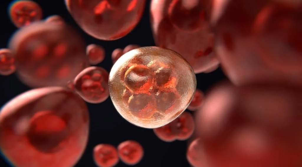
Liver cancer remains one of the deadliest cancers worldwide, largely because it is often diagnosed at an advanced stage when treatment options become limited. Now, a new microbubbles for liver cancer study from China suggests that an innovative ultrasound-assisted approach could make existing therapies more effective.
Published in the peer-reviewed journal Scientific Reports, the research found that combining microscopic gas-filled bubbles with ultrasound helped shrink tumors more effectively than standard treatment alone in patients with an aggressive form of liver cancer. While the findings are still early and require larger clinical trials, they point to a promising new direction for localized cancer treatment.
For patients, doctors, and researchers, the study highlights how a technology originally designed for medical imaging may have a much bigger role to play in cancer care.
TL;DR
- Researchers in China tested ultrasound-assisted microbubble therapy in patients with advanced liver cancer.
- Patients receiving the experimental treatment experienced greater tumor shrinkage than those receiving standard therapy alone.
- Microbubbles are tiny gas-filled spheres that respond to ultrasound waves.
- The technique may improve how cancer therapies reach tumors without replacing existing treatments.
- More large-scale clinical studies are needed before the treatment becomes standard medical practice.
What are microbubbles for liver cancer?
Microbubbles are microscopic spheres filled with gas and surrounded by a thin shell made of lipids or proteins. They were originally developed as contrast agents to improve ultrasound imaging by making blood vessels easier to visualize.
Over the past decade, researchers have discovered that these tiny bubbles can do much more than improve medical scans.
When exposed to carefully controlled ultrasound waves, microbubbles rapidly expand and contract. This mechanical activity can temporarily increase the permeability of nearby blood vessels and cell membranes, potentially allowing medications to penetrate tumors more effectively.
Rather than acting as a cancer drug themselves, microbubbles function as treatment enhancers, helping existing therapies reach their target more efficiently.
How did the new liver cancer study work?
Researchers compared two patient groups
Scientists from the Affiliated Hospital of Guangdong Medical University and Huzhou Central Hospital evaluated two groups of patients with an aggressive form of liver cancer.
One group received conventional treatment, while the second group received the same therapy combined with ultrasound-assisted microbubble treatment.
The comparison allowed researchers to determine whether adding microbubbles could improve treatment effectiveness beyond current medical approaches.
The experimental group showed better tumor shrinkage
According to the study, tumors shrank more in patients who received the combination treatment than in those treated with standard therapy alone.
The findings suggest that ultrasound-triggered microbubbles may improve how effectively treatment reaches cancer tissue.
Importantly, the study focused on improving existing therapies—not replacing them.
How do ultrasound and microbubbles work together?
The concept is surprisingly straightforward.
After being injected into the bloodstream, microbubbles circulate through blood vessels, including those supplying tumors.
When physicians direct ultrasound waves at the treatment area:
- The microbubbles begin expanding and contracting rapidly.
- Their movement creates mechanical effects around nearby cells.
- Blood vessel walls may become temporarily more permeable.
- Cancer treatments can potentially penetrate deeper into tumor tissue.
- Drug delivery may become more targeted while limiting effects on surrounding healthy tissue.
Researchers sometimes describe this process as temporarily opening a “window” that allows therapies to reach areas they might otherwise struggle to access.
Consider adding an infographic here illustrating how microbubbles travel through blood vessels and respond to ultrasound before helping deliver treatment into tumor tissue.
Why does this matter for liver cancer treatment?
Liver cancer is particularly difficult to treat because many tumors develop in patients whose liver function is already compromised by chronic disease or cirrhosis.
Advanced-stage tumors often respond poorly to treatment due to several challenges:
- Limited drug penetration into tumor tissue.
- Complex blood vessel networks.
- Resistance to conventional therapies.
- Risk of damaging healthy liver tissue.
If microbubble-assisted ultrasound can improve how treatments reach cancer cells, physicians may eventually achieve better outcomes without dramatically increasing medication doses.
That could potentially reduce side effects while improving treatment effectiveness.
However, the current evidence remains preliminary, and researchers caution against viewing the technology as a cure.
What makes microbubbles unique?
Microbubbles stand out because they combine diagnostic and therapeutic capabilities.
Originally developed for imaging, they are now being investigated across several medical applications.
Potential uses include:
- Improving targeted drug delivery.
- Enhancing localized cancer treatment.
- Increasing precision in ultrasound-guided therapies.
- Supporting research into non-invasive treatment techniques.
Outside medicine, microbubbles are also used in:
- Environmental cleanup.
- Industrial engineering.
- Water treatment technologies.
- Chemical processing.
Their ability to respond predictably to ultrasound makes them highly versatile across multiple scientific disciplines.
What are the limitations of the research?
Although the findings are encouraging, several important questions remain unanswered.
The study is still early
The published research represents an important step, but not definitive proof that the treatment should become routine clinical practice.
Future studies will need to determine:
- Whether the results can be replicated in larger patient populations.
- Which liver cancer patients benefit the most.
- The long-term survival outcomes.
- Potential side effects after repeated treatments.
- The optimal ultrasound settings and treatment schedules.
Tumor shrinkage is only one measure
Reducing tumor size is an encouraging outcome, but cancer researchers also evaluate:
- Overall survival.
- Progression-free survival.
- Quality of life.
- Recurrence rates.
- Long-term safety.
Only additional clinical trials can determine whether improved tumor shrinkage translates into longer survival.
Could this technology be used for other cancers?
Potentially, yes.
Researchers worldwide are already studying ultrasound-responsive microbubbles for several types of cancer, including:
- Breast cancer.
- Pancreatic cancer.
- Brain tumors.
- Prostate cancer.
- Ovarian cancer.
The same principle of improving localized drug delivery could apply to many solid tumors where medications struggle to penetrate effectively.
However, each cancer behaves differently, meaning separate clinical trials will be necessary before broader adoption.
What happens next?
The next phase of research will likely involve larger multicenter clinical trials designed to answer several critical questions.
Scientists will need to establish:
- Which patients benefit the most.
- Whether the treatment improves survival.
- How frequently therapy should be administered.
- Whether it works alongside newer targeted drugs and immunotherapies.
- Whether the approach remains safe over longer follow-up periods.
If future research confirms the current findings, microbubble-assisted ultrasound could eventually become an important addition to existing liver cancer treatment strategies rather than replacing current therapies.
Why this study deserves attention
Medical innovation often comes from improving existing treatments instead of inventing entirely new drugs.
This research illustrates that principle well.
By using microscopic gas bubbles and ultrasound—technologies already familiar to modern medicine—researchers may have found a way to help established cancer treatments work more effectively.
While it is far too early to describe the approach as a breakthrough or cure, the study offers a promising example of how engineering, imaging technology, and oncology can intersect to create smarter, more targeted cancer therapies.
For patients facing advanced liver cancer, even incremental improvements in treatment effectiveness could have meaningful clinical significance if confirmed through future research.