
A 60-year-old man in Spain arrived at a hospital with severe headaches and changes in his behavior, symptoms that initially led doctors to suspect brain cancer. Instead, advanced imaging revealed something far more unusual: tapeworm larvae lodged inside his brain.
The rare medical case, published in the journal Emerging Infectious Diseases, has drawn attention because the patient had no recent travel history and no obvious risk factors. The discovery highlights how parasitic infections can sometimes imitate life-threatening conditions such as brain tumors and why physicians should keep rare diseases in mind, even in countries where they are uncommon.
What happened to the Spanish man?
The patient sought medical attention after suffering from severe headaches for nearly two weeks. Family members also noticed changes in his behavior, prompting doctors to perform neurological examinations.
Apart from slightly slowed movements, his neurological examination was largely normal. Blood tests also appeared unremarkable except for elevated Immunoglobulin E (IgE), an antibody that often increases during allergic reactions or parasitic infections.
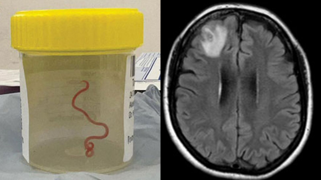
A CT scan painted a more alarming picture.
Doctors found multiple brain lesions surrounded by swelling, a pattern commonly associated with metastatic cancer that has spread to the brain.
Because the patient had no weakened immune system and had never traveled abroad, cancer initially appeared to be the most likely diagnosis.
How doctors discovered the real cause
To identify the suspected cancer, physicians ordered an extensive series of tests, including:
- Whole-body contrast-enhanced CT scan
- Colonoscopy
- PET/CT imaging
- Additional laboratory tests
Surprisingly, none of these examinations found any evidence of cancer.
The breakthrough came when doctors performed an MRI scan.
Unlike the earlier CT images, the MRI clearly showed tiny encapsulated larvae with visible tapeworm heads inside the brain lesions. Instead of tumors, the patient had neurocysticercosis, a parasitic infection caused by the pork tapeworm.
The finding completely changed the diagnosis and treatment strategy.
What is neurocysticercosis?
A parasitic infection that affects the brain
Neurocysticercosis (NCC) occurs when larvae of the pork tapeworm (Taenia solium) invade the central nervous system.
Rather than remaining in the intestines as adult worms, the parasite’s larvae migrate through the bloodstream and become trapped in tissues, including the brain, where they form fluid-filled cysts.
These cysts may remain unnoticed for years before triggering symptoms.
Common symptoms
Symptoms vary depending on the number and location of cysts but can include:
- Persistent headaches
- Seizures
- Memory or behavioral changes
- Confusion
- Difficulty with movement or coordination
- Increased pressure inside the skull
Because these symptoms overlap with brain tumors, strokes, and other neurological disorders, diagnosis can be challenging.
How do tapeworms spread?
People often associate tapeworm infections with eating undercooked pork, but neurocysticercosis usually develops differently.
There are two primary routes of infection:
Eating undercooked infected pork
When someone eats pork containing tapeworm larvae, the parasites mature into adult worms inside the intestines.
Swallowing tapeworm eggs
A far more dangerous route occurs when microscopic tapeworm eggs enter the body through food, water, or surfaces contaminated with human feces.
Once swallowed, the eggs hatch, penetrate the intestinal wall, enter the bloodstream, and travel to organs such as the brain, muscles, eyes, and skin, where they develop into cysts.
This second route causes neurocysticercosis.
Why this case surprised researchers
Spain is not considered a country where Taenia solium is commonly found, making the diagnosis especially unexpected.
Researchers noted that the patient had:
- Never traveled internationally
- No significant immune disorder
- No obvious exposure to endemic regions
Investigators suggested one possible explanation.
The man had previously worked in construction, where several coworkers came from countries in which pork tapeworm infections are more common. Researchers believe he may have unknowingly been exposed through shared meals or contaminated restroom facilities if one coworker carried the parasite.
However, they stressed that this remains only a hypothesis.
The exact source of the infection has never been confirmed.
Why doctors initially suspected cancer
Brain imaging can sometimes make parasitic infections look remarkably similar to metastatic cancer.
Both conditions may produce:
- Multiple brain lesions
- Swelling around affected tissue
- Neurological symptoms
- Abnormal imaging findings
Only high-resolution MRI imaging revealed the distinctive appearance of tapeworm larvae, allowing doctors to distinguish the infection from malignant tumors.
The case illustrates why imaging findings alone may not always provide a definitive diagnosis.
Why this case matters for medicine
Researchers say the report serves as an important reminder that rare diseases can appear even in places where they are not commonly diagnosed.
Modern migration, international travel, and global workforces mean physicians increasingly encounter illnesses traditionally associated with other parts of the world.
The study’s authors emphasized that doctors should include neurocysticercosis among possible diagnoses when patients present with multiple brain lesions, even if they have no travel history.
Making the correct diagnosis can prevent unnecessary cancer treatments while ensuring patients receive appropriate therapy for parasitic infections.
Can neurocysticercosis be prevented?
Although uncommon in many developed countries, experts recommend several preventive measures:
- Cook pork thoroughly before eating.
- Wash hands carefully after using the restroom.
- Practice proper food hygiene.
- Drink clean, safe water.
- Improve sanitation to prevent fecal contamination.
- Seek treatment for intestinal tapeworm infections to stop further transmission.
Public health experts note that improved sanitation and food safety remain the most effective long-term strategies for reducing infections worldwide.
The bigger takeaway
This unusual Spanish case demonstrates how medicine can still surprise even experienced clinicians. What initially appeared to be metastatic brain cancer ultimately turned out to be a rare parasitic infection that mimicked a tumor almost perfectly.
For researchers, it reinforces the importance of considering uncommon diagnoses even when patients lack traditional risk factors. For the public, it serves as a reminder that maintaining good hygiene, proper food preparation, and awareness of parasitic diseases remain important, even in countries where such infections are considered rare.
TL;DR
- A 60-year-old Spanish man developed headaches and behavioral changes.
- Initial scans suggested metastatic brain cancer.
- An MRI later revealed tapeworm larvae inside his brain.
- Doctors diagnosed neurocysticercosis, a parasitic infection caused by Taenia solium.
- The patient had no travel history, making the source of infection difficult to identify.
- Researchers say the case reminds doctors not to rule out rare parasitic infections based solely on geography.